Zygomatic Classical Technique or Anatomy-Guided Approach?
For several decades, zygomatic implantation has been the preferred technique for patients with either maxillectomy defects or the atrophic edentulous maxilla. Yet, up until recently, the original Brånemark protocol – utilizing the intra-sinus pathway – was the only documented way of fully rehabilitating patients. In patients with pronounced buccal concavities, however, this approach often resulted in the palatal emergence of the implant head. This meant that some experienced discomfort, including chronic rhinosinusitis or sinus congestion.
"96,8% success rate of zygomatic implants"

Due to these concerns, several changes to the Brånemark technique have been documented and indeed, the possibility of incorporating an extra-sinus approach has been proposed by various clinicians. This approach allows the placement of the head to be aligned on or near the top of the residual crest, by-passing the maxillary sinus and as research shows, results in a better outcome for the patient.
Exploring the ZAGA approach
As a refinement to the extra-sinus technique, Aparicio first described the concept of the Zygomatic Anatomy-Guided Approach—otherwise known as the ‘ZAGA’ technique. In this case, any placement of zygomatic implants is guided by a prosthetically and anatomically driven approach and applies to not only patients with buccal concavities but all types of maxillary anatomies.
Having the flexibility to place either all or part of the body of the zygomatic implant outside of the maxillary sinus it was thought to result in fewer complications due to the following reasons:
- A smaller part of the implant may be inside the sinus cavity
- Implants placed in this way are guided by crest position, so abnormal communication between the maxillary sinus and the oral cavity is less likely.
In addition, it was suggested that a good understanding of the ZAGA concept would help the clinician to make better use of the crestal bone to ensure:
- Better bone integration at the neck and body of the implant and
- Greater soft tissue control to ensure better coverage of the zygomatic implant

Diagnosing CRS
Of course, when dealing with zygomatic implant problems such as chronic rhinosinusitis (CRS), they first need to be correctly identified. Surprisingly, there are no specific documented dental protocols for identifying and diagnosing CRS.
Ear Nose & Throat (ENT) literature, for example, states that two or more symptoms need to be present in order to diagnose chronic rhinosinusitis (CRS) fully. In addition, definitive findings are usually carried out utilizing a nasal endoscopy and/or a CT scan. Yet, in most of the studies involving zygomatic implants, the term used to describe any form of sinus pathology is simply ‘sinusitis.’ Clearly, this doesn’t clarify or specify the problem type. To provide greater clarity, a study was set up with two main aims. The first aim was to compare the classical zygomatic technique (CZT) to those patients treated using the ZAGA approach. Where factors such as:
- Long-term survival outcomes
- Implant stability
- Sinus conditions
- Soft tissue sealing and
- Prosthesis design
were all taken into account. The second aim of the study was to create and propose a standardized system for reporting rhinosinusitis.
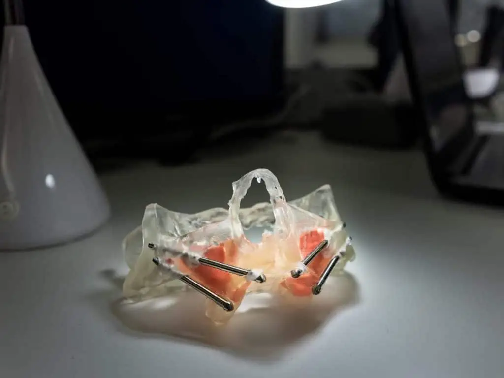
The Implant Placement
Between 1998 and 2002, all 172 implants were placed by a single dental surgeon. All were machined-surface titanium implants manufactured by Nobel Biocare. The 131 regular implants were sized between 7mm and 18mm and the remaining 41 zygomatic implants ranged from 30-50mm in length. In total 55 regular implants were anchored in the canine areas, 29 other regular implants were split between the Pterygoid and the Pyramidal processes, and the remaining 41 zygomatic implants were anchored into the zygomatic (cheek) bone.
Study Background

Firstly a control group was set up which included 22 patients with atrophic edentulous maxilla. Regular and zygomatic implants were placed using the classical zygomatic technique (CZT). A further 80 patients were also treated with implants using the principles of the ZAGA approach. These patients were already undergoing a periodic maintenance program and were incorporated into the study as the test group.
The surgical period lasted from January 2004 to October 2009, with an average follow-up of 4.62 years. In addition, all patients included in the test group had 3 years of prosthetic follow-up. Finally, at the end of the study, all patients were contacted and underwent a full and final clinical and radiological evaluation. They were also invited to answer two specific questions on the following:
- Health and status of their sinuses and implants, respectively, and…
- Degree of satisfaction throughout the course of the treatment.
Implants Used
In all, 857 titanium implants were placed by a single surgeon. 660 were regular implants and the remaining 197 were zygomatic implants. In both control and test groups, regular implants (RI’s) were anchored into a variety of places including residual bone in the canine areas, the sub-nasal crest, the sphenoid bone, the anterior nasal spine, and the palatine bone. In the control group, all zygomatic implants were placed as per the original technique. Whereas, in the test group, all ZI’s were placed using the modified ‘ZAGA’ approach.
The Method
Depending upon the anatomical structure of each patient’s maxilla, the zygomatic implant pathway was divided into 5 categories and in all ZAGA cases – irrespective of structure group – a one-stage, immediate loading procedure with abutment connection and temporary prosthesis was used. In all CZT cases, a 2 stage procedure with 5-6 months of healing time was undertaken.
Regular periotest measurements were also undertaken in both groups to access implant stability and a sinus symptomatology questionnaire was undertaken to identify the presence (if any) of sinusitis symptoms. In addition, CBCT scans were used on 102 patients and scored/ranked using the Lund-Mackay (LM) score.
The Results
After extensive testing, it was found that both classical and ZAGA procedures were found to have similar positive clinical outcomes with regards to implant survival. However, the ZAGA concept was shown to minimize the risk of maxillary sinus problems whilst quickly rehabilitating severely atrophic maxillae. Moreover, results from the questionnaire showed that Zygomatic implants placed in this way are less visible, less bulky, and more comfortable for the patient. Overall, this study shows that the ZAGA concept works in terms of minimizing risk and obtaining long-lasting comfort for the patient. Therefore strong evidence would suggest that it should be considered as an integral protocol when evaluating best patient outcomes for those with atrophic maxilla in the future.