- Katarina

2005: the first externalized zygomatic implants, presented at EuroPerio Madrid
Twenty years ago, at EuroPerio in Madrid, the team at Clínica Aparicio presented something that was not in any protocol at the time: zygomatic implants placed along the outside of the maxillary wall instead of through the sinus. Drs. Wafaa Ouazzani, Stefan Lundgren, Lars Sennerby and Carlos Aparicio showed one year of clinical results at the EuroPerio conference. The paper followed in 2006 in the Journal of Clinical Periodontology (1).
Until then everyone followed the original surgical technique (OST) described by Professor P.I. Brånemark: the implant enters the sinus and exits through the zygoma. It worked, but two problems kept coming back: sinus infections, and a prosthesis that sat too far palatally and felt bulky. Moving the implant outside the sinus wall, when the patient’s anatomy called for it, was the first time the surgery was adapted to the patient rather than the patient to the surgery. That decision is where the ZAGA Concept starts.
Dates for the upcoming zygomatic implant courses are published on the course page.

The same finding in Brazil
Dr. Migliorança’s group in Brazil reached a similar conclusion at about the same time, working independently. Two teams on two continents arriving at the same answer is a good sign the answer is about anatomy and not about one surgeon’s preference.
Zygomatic implants were already the option for patients without enough bone for conventional implants. What the externalized path changed was the complication profile: fewer sinus problems, and a prosthesis that could sit where the teeth used to be.
From a technique to a classification: ZAGA 0 to ZAGA 4
The three-year prospective study on extra-maxillary placement, published online in 2008 in Clinical Implant Dentistry and Related Research (2), is where the Zygoma Anatomy-Guided Approach took shape. Instead of one trajectory for everyone, the approach reads the shape of the zygomatic buttress and the alveolar crest and sorts patients into five types, ZAGA 0 to ZAGA 4. Each type implies a different implant path and a different amount of bone contact, and that in turn decides how the soft tissue and the prosthesis will behave.
The complications the classification is designed to avoid are the ones every zygomatic surgeon has seen:
- soft tissue dehiscence around the implant
- sinusitis
- a bulky, palatal prosthesis

What anatomy-guided means in practice
A standardized technique treats a ZAGA 0 maxilla, where the implant can run inside the sinus with its head on the crest, the same as a ZAGA 4, where the crest is almost gone and the implant has to run outside the maxilla under the mucosa. That is the error the approach corrects. In practice it means:
- the trajectory is planned on the patient’s anatomy, not chosen by habit, which is what lowers the complication rate
- soft tissue over the implant body is part of the plan, so healing and the look of the final prosthesis are more predictable
- the implant head ends up under the prosthetic tooth, which keeps the prosthesis slim and the surrounding structures intact over the long term

Twenty years on: the implants caught up with the approach
For most of those twenty years the approach was applied with implants designed for the original intra-sinus path. That changed with the ZAGA Round and ZAGA Flat implants, developed with Straumann. Round is for the ZAGA types where the implant body sits in bone. Flat, with its flat side against the maxillary wall, is for the types where the body runs outside the maxilla, so it sits closer to the bone and leaves less of a bulge under the mucosa. Matching the implant shape to the ZAGA type is the latest step in the direction the 2005 presentation set: the anatomy decides, and the hardware follows.
Patients who twenty years ago were told there was nothing to be done are now routinely rehabilitated with a fixed prosthesis, without grafting. That is the practical result of the whole story.
The ZAGA Centers network
The approach spread through courses and through the surgeons who adopted it, and that is how ZAGA Centers came about: a network of clinics whose surgeons are trained and certified in the ZAGA Concept, so that a patient with a severely atrophic maxilla can find, in their own country, someone who plans zygomatic cases this way. The centers also feed cases and data back into the research, which is how the classification keeps being tested. The map below shows the network as of May 2025.

What comes next
Planning is now digital in most ZAGA Centers: CBCT-based planning, guided surgery in selected cases, and 3D-printed patient models to rehearse the surgery before the patient is in the chair. None of it changes the principle. It makes reading the anatomy faster and the trajectory more repeatable.
Twenty years after Madrid, the shift from one technique for everyone to a trajectory chosen per patient is the standard we teach, publish and certify against. Thank you to everyone who has placed, studied or taught these implants with us since 2005.
References:
Read more
The science page goes further into the ZAGA classification, the published studies and the Straumann ZAGA implants: ZAGA Science.

Past event
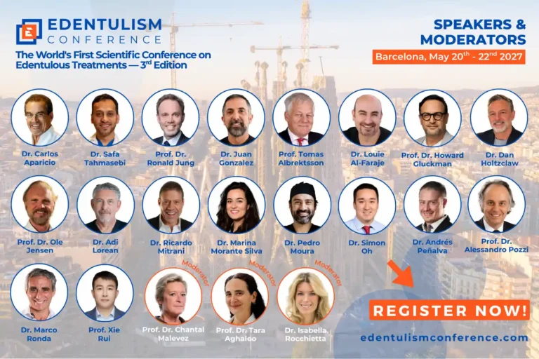
The twentieth anniversary year also had its conference. Edentulism 2025 took place in Barcelona from 5 to 7 June 2025, with Prof. Tomas Albrektsson, Dr. Carlos Aparicio, Dr. Taras Yurov and a programme built around full-arch rehabilitation and the treatment of edentulous patients. A special edition of the ZAGA Course ran in the days before it (2 to 4 June), so the week covered zygomatic, pterygoid and transnasal implants from lecture to hands-on.
The next edition is Edentulism 2027. Dates, speakers and registration are published at edentulismconference.com.
- Download the brochure